| Abstract |
Extratesticular localization of neurofibroma have been reported extremely rarely in the literature. We are reporting such rare case with a different presentation. A 60-year-old male patient reported twith a swelling in the scrotum, diagnosed as hydrocele and was operated. Later on, patient has developed scrotal thickness and ulcer. Edge biopsy was taken and to our surprise, on histopathology, it came as neurofibroma of the scrotal skin.
Keywords: Extratesticular, hydrocele, neurofibroma, scrotum, skin, ulcer
| How to cite this article: Singal R, Singal RP, Singal S, Sekhon A. Neurofibroma of the scrotum: An unbelievable experience. Ann Trop Med Public Health 2012;5:370-2 |
| How to cite this URL: Singal R, Singal RP, Singal S, Sekhon A. Neurofibroma of the scrotum: An unbelievable experience. Ann Trop Med Public Health [serial online] 2012 [cited 2020 Aug 15];5:370-2. Available from: https://www.atmph.org/text.asp?2012/5/4/370/102063 |
| Introduction |
Neurofibroma (NF) is a benign tumor of the nerve sheath, which is considered to originate from the Schwann cell. [1] Since neural tissues are found throughout the body, these tumors can occur in a variety of sites (anywhere within the central or peripheral nervous system), especially in the neck, thorax, cranium, retroperitoneum, and flexor surfaces of the extremities but localization within the scrotum is extremely rare. [2],[3],[4]
| Case Report |
A 60-year-old male presented with a 1-year history of a painless, slowly growing, left-sided scrotal mass. There was no history of surgical or medical problems, and there was no significant family history.
On examination- the mass was located in the left side of the testis. It measured 10 cm x 20 cm. It was oval in shape, non-tender, firm in consistency, and including the testis and transillumination test was present. It was not attached to the scrotal skin or other underlying structures. There were no palpable inguinal lymph nodes.
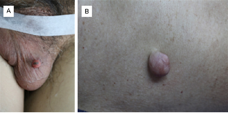
Ultrasonography (USG) showed an oval, well defined, soft tissue lesion diagnosed as hydrocele on left side. Surgical exploration was done for hydrocele and later on he developed scrotal thickness and ulcer [Figure 1]. Then we excised the skin with proper margins and sent for histopathology. Margins were freshened with primary closure of the wound was done. It healed very well. To our surprise,on histopathology diagnosis came as neurofibroma of the scrotum. Microscopically, it was showing proliferation of haphazardly arranged neural tissue growing beneath a flattened epidermis [Figure 2] and [Figure 3].
 |
Figure 1: Post-operative picture showing ulcer on right side of scrotal region |
 |
Figure 2: Photomicrograph showing proliferation of haphazardly arranged neural tissue growing beneath a flattened epidermis (H and E, ×40) |
| Figure 3: Photomicrograph of the tumor showing fascicular growth pattern and serpentine nuclei (H and E, ×200)
Click here to view |
| Discussion |
Peripheral nerve sheath tumors arise from the neural crest. In the male population, benign intrascrotal lesions are commonly seen. [5] The first case of solitary neurofibroma in the scrotum was described by the Yamamoto et al, [6] Neurofibroma can be solitary or multiple and are commonly seen in between of age 8 and 77 years. Most of them occur in paratesticular tissue such as epididymis or spermatic cord. Neurofibromas are well-differentiated nerve sheath tumors of benign origin, which commonly present as a part of systemic disease NF-1 or NF-2. [7] Unlike testicular lesions, which are 95% malignant, paratesticular lesions are mostly benign. Usually, surgical exploration is required to rule out intrascrotal malignant processes. [5]
Benign tumors of the scrotum are leiomyomas, lipomas, fibromas, hemangiomas, and epidermoid cysts have been described as. Solitary neurofibroma within the scrotum, which is unassociated with neurofibromatosis, is an extremely rare benign tumor. [8] NF tumors are not encapsulated and have a softer consistency. Microscopically, these are formed by combined proliferation of all the elements of peripheral nerve axons, Schwann’s cells, fibroblasts, and perineural cells. Schwann’s cells are usually the predominant cellular elements in the tumor. [5]
These tumors may originate anatomically from the testis, tunics, and subcutaneous neural tissue. In the literature, eight cases of solitary neurofibroma were reported previously, which involved the external genitalia in the absence of von Recklinghausen’s disease. [3],[4] We recognized that this mass was separate from the testis, vas deferens, and epididimis. In this case, we agree with Issa et al,[9] that the tumor must have originated from the genital branch of the genitofemoral nerve lying posteriorly to the spermatic cord. This nerve innervates cremasteric muscle and distributes branches to the skin of the scrotum and adjacent thigh.
A similar report has also been published by Milathianakis et al,[3] Although treatment for most primary tumors has historically been radical inguinal orchidectomy, most benign tumors can now be managed by testis sparing surgery. [10] For this reason, in neurofibroma, the treatment is surgical excision of tumor. Harding et al,[11] reported that 10.4% of men with testicular and paratesticular tumors underwent a scrotal orchidectomy or had a scrotal incision before an inguinal orchidectomy. In their study, the authors suggested that scrotal incision is unlikely to affect the risk of loco-regional recurrence. These tumors have an excellent prognosis, and they can be cured by complete surgical excision. [8]
| Conclusion |
Solitary neurofibroma are rare entity especially in scrotal region. Frozen section microscopic examination should be performed pre-operatively to ascertain whether the tumor is benign or malignant in case of ulcer. Treatment of choice is surgical approach with complete excision of the mass. Although an extremely rare disease, neurofibroma should be considered in the differential diagnosis of testicular and paratesticular tumors.
| References |
| 1. | Mishra VC, Kumar R, Cooksey G. Intrascrotal neurofibroma. Scand J Urol Nephrol 2002;36:385-6. |
| 2. | Trovarelli S, Tallis V, Tripodi S, Miracco C, Ponchietti R. Extratesticular intraescrotal neurofibroma: Case report. Arch Esp Urol 2006;59:905-8. |
| 3. | Milathianakis KN, Karamanolakis DK, Mpogdanos IM, Trihia-Spyrou EI. Solitary neurofibroma of the spermatic cord. Urol Int 2004;72:271-4. |
| 4. | Rosai J. Soft tissues. Ackerman’s Surgical Pathology. 8 th ed. New York: Mosby Year Book; 1995. p. 2041-2. |
| 5. | Rubenstein RA, Dogra VS, Seftel AD, Resnick MI. Benign intrascrotal lesions. J Urol 2004;171:1765-72. |
| 6. | Yamamoto M, Miyake K, Mitsuya H. Intrascrotal extratesticular neurofibroma. Urol 1982;20:200-1. |
| 7. | Dwivedi S, Baisakhiya N, Bhake A, Bhatt M, Agrawal A. Giant solitary neurofibroma presenting as a neck mass in an infant. J Neurosci Rural Pract 2010;1:32-4. |
| 8. | Turkyilmaz Z, Sonmez K, Karabulut R. A childhood case of intrascrotal neurofibroma with a brief review of the literature. J Pediatr Surg 2004;39:1261-3. |
| 9. | Issa MM, Yagol R, Tsang D. Intrascrotal neurofibromas. Urol 1993;41:350-2. |
| 10. | Agarwal PK, Palmer JS. Testicular and paratesticular neoplasms in prepubertal males. J Urol 2006;176:875-81. |
| 11. | Harding M, Paul J, Kaye SB. Does delayed diagnosis or scrotal incision affect outcome for men with nonseminomatous germ cell tumours? Br J Urol 1995;76:491-4. |
Source of Support: None, Conflict of Interest: None
| Check |
DOI: 10.4103/1755-6783.102063
| Figures |
[Figure 1], [Figure 2], [Figure 3]


