| Abstract |
Myiasis is a condition resulting from the invasion of living human and animal’s tissues by larval stages of flies. study the cutaneous and genito-urinary myiasis in the Republic of Yemen. Two females and one male, Yemeni patient 12-30 years old presented with urethra-genital discharge, burning, and difficulty during micturition (dysuria). They saw whitish brownish blackish like worms passed in their urine. Three males, Yemeni patients 2 months to 30 years old had solitary and multiple furuncle like skin lesions in upper left thigh (myiasis pubis), right buttock (myiasis neonatorum) and in the upper trunk. It was associated with stabbing pain (caused by creeping a foreign body subcutaneously). The duration varied from 2 days to 1 week. The larvae were detected in the urine and the skin lesions. Swab from the urethral discharge was not specific. I used the forceps accidentally and gently caught the larvae and removed them from the fruncular skin lesions. The clinical data and detection of the larvae showed the two females and one male case were genito-urinary myiasis, which caused by latrine Fannia fly larvae during defecation or sleeping and ended spontaneously. The three male patients had cutaneous myiasis, and the causative agent was the larvae of the tumbu fly, Cordylobia anthropophaga species. It penetrates the patients’ normal skin and removed by forceps or squeezing the skin lesion by the patient himself thinking that is abscess to remove the pus. Cutaneous and genito-urinary myiasis was not rare in the Republic of Yemen. Health education, hygiene, and control the flies restrict the incidence of this disease.
Keywords: Al-Kuwait University Hospital, Sana′a University, Medical School, Department of Dermatology, Myiasis, cutaneous, genito urinary
| How to cite this article: Al-Malmi MA. Myiasis in Republic of Yemen (cutaneous myiasis three cases reported and genito-urinary myiasis three cases reported). Ann Trop Med Public Health 2014;7:238-43 |
| How to cite this URL: Al-Malmi MA. Myiasis in Republic of Yemen (cutaneous myiasis three cases reported and genito-urinary myiasis three cases reported). Ann Trop Med Public Health [serial online] 2014 [cited 2020 Aug 10];7:238-43. Available from: https://www.atmph.org/text.asp?2014/7/5/238/151778 |
| Introduction |
Myiasis is the term applied to the infestation of live humans and vertebrate animals with the larvae (maggots) of Diptera (two-winged) flies. In humans, infestation may affect the skin, wounds, intestines, and body cavities (oral, nasal, aural, ocular, sinusal, intestinal, and vaginal and urethra). [1],[2],[3],[4],[5],[6],[7],[8],[9],[10],[11] When open wounds are involved, the myiasis is known as traumatic, and when boil-like, the lesion is termed furuncular. Cordylobia anthropophaga (also referred to as “tumbu fly”, “mango fly”, “skin maggot fly” or “verde cayor”) is endemic in tropical Africa. [2],[3],[4],[5],[6],[7],[8],[9],[10],[11],[12],[13] Furuncular myiasis as a result of C. anthropophaga infestation has been endemic in the West African subregion for >135 years. [3],[4],[5],[6],[7],[8],[9],[10],[11],[12],[13],[14],[15] The other flies that cause furuncular myiasis include Cordylobia rhodaini (Lund fly, found in the rainforest areas of tropical Africa) and Dermatobia hominis (human botfly, which is endemic in Central and South America). [2],[6],[7],[8],[9],[10],[11],[12],[13],[14],[15],[16],[17],[18],[19] The mode of transmission of D. hominis differs from the Cordylobia species. The eggs of D. hominis are carried to the host by a blood-sucking insect, such as mosquitoes and ticks and the hatched larvae invade exposed skin of the trunks, head, and limbs. The eggs of Cordylobia species are, however, deposited on the soil or wet and soiled clothes hung outside for drying. The hatched larvae invade unexposed skin (of the buttocks, trunk, the limbs, and penis) in contact with the wet clothes. [1],[2],[3],[4],[5],[6],[7],[8],[9],[10],[11],[12],[13],[14],[15],[16],[17],[18],[19],[20],[21],[22],[23],[24],[25],[26],[27],[28],[29]
| Case Reports |
Case 1
A 33-year-old Yemeni soldier working in Yemen Army in Safer Mareb about 200 km east capital Sana’a city. He presented to the Al-Kuwait University Hospital skin clinic in summer June 1998. He was very irritable and nervous, and he felt with severe acute stabbing pain in multiple furuncular skin lesions in the upper trunk or chest and upper back. His right breast was involved with one furunuclar skin lesion. With my experience, I suspected that the patient had multiple furuncular myiasis [Figure 1] and [Figure 2]. Without local anesthesia, I admitted him very quick to the minor surgical operation room. I directly and very gently used the forceps and catch the whitish alive larvae one by one from all the furuncles [Figure 3], [Figure 4] and [Figure 5]. I did not request for this patient any either laboratory or imaging investigations. I wrote for him topical and systemic antibiotic to protect him against the secondary infection occurrence.
| Figure 1: Acute multiple large furuncles skin lesions in the upper trunk or chest without discharge pus or sinuses. Dermotrobia hominis larvae were seen after removal
Click here to view |
| Figure 2: The large acute furuncle in the right breast without pus or sinuses
Click here to view |
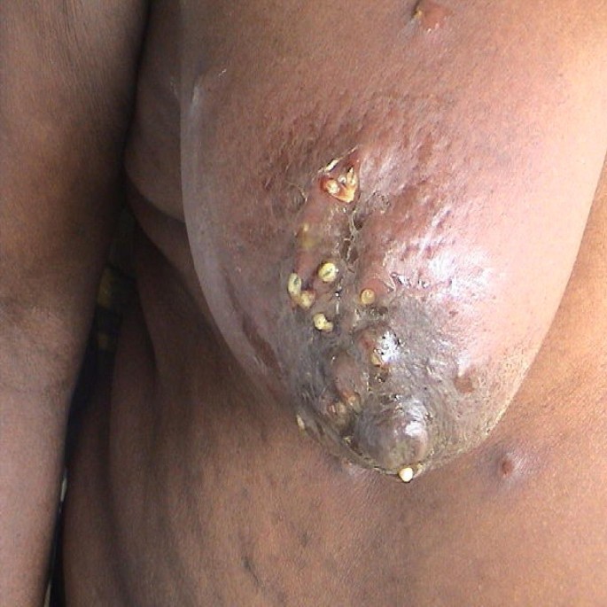 |
Figure 3: Large acute furuncles skin lesion in the left upper chest Demetrobia hominis larva seen after removal
Click here to view |
| Figure 4: Large acute furuncle skin lesion in the upper right chest Demetrobia hominis was seen after removal
Click here to view |
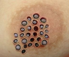 |
Figure 5: Large acute furuncle skin lesion in the right upper back Demetrobia hominis larva was seen after removal
Click here to view |
Case 2
A 23-year-old male farmer Yemeni patient came to my private clinic in May 1993 from Amran 50 km outside Sana’a city. He had solitary boil or furuncle like skin eruption in the upper left thigh of day’s duration [Figure 6]. He said he squeezed this lesion by his fingers than one worm come outside. He was afraid and annoyed if more worm inside his skin lesion. I calmed him, that is, while the worm or larva removed by him squeezing, he will be cured. He must take care from any other flies in his home in the village, and he may get a new reinfection. I wrote for him topical and systemic antibiotics.
| Figure 6: Solitary large furuncle skin lesion in the upper right thigh
Click here to view |
Case 3
A 2-month-old Yemeni boy baby presented with solitary furuncle like skin lesion in his left buttock. Mother brings here in a small glass box, dead larva. She said she squeezed the lesion by her fingers, because she thought that is obsess to get rid of the pus. The larva comes out. Show the small yellowish whitish larva beside the skin lesion after removal by squeezing here mother fingers [Figure 7]. The baby treated by topical and systemic antibiotics. I calmed here mother that is cutaneous myiasis and take care the hygiene of some flies.
| Figure 7: Large acute furuncle skin lesion in buttock of neonate boy
Click here to view |
Cases 4-6
Two adult females and one adult male, Yemeni farmers presented with dysuria. They showed some small worms like passed through their urethra openings. There was no urethral obstruction. The larvae passed spontaneously with the urine and cured completely [Figure 8], [Figure 9], [Figure 10], [Figure 11] and [Figure 12].
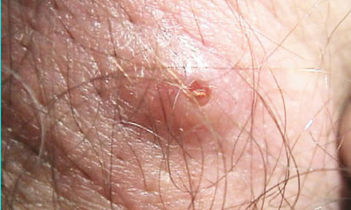 |
Figure 8: Large larva of Demetrobia hominis causes cutaneous myiasis
Click here to view |
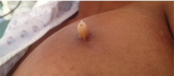 |
Figure 9: Larvae of latrine Fannia fly species causes of ginito urinary myiasis
Click here to view |
 |
Figure 10: High magnification of latrine Fannia fly larvae: Ventral view
Click here to view |
| Figure 11: Latrine Fannia fly larvae: Dorsal view
Click here to view |
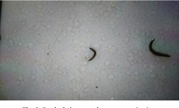 |
Figure 12: Other species of Latrine Fannia fly larvae after removal from urethera
Click here to view |
Comment
The adult fly resembles a bumblebee (see image below); it is short lived and survives for little more than a week. It does not feed and is infrequently seen. The life cycle of the botfly is unique, as the female, egg-bearing fly attaches her eggs to the abdomen of a blood-sucking arthropod (means of transportation known as phoresy), usually a mosquito (although 40 other species of insects and ticks have been reported). When the mosquito takes a blood meal from a warm-blooded animal, the local heat induces the eggs to hatch and drops to the skin of the host and enters painlessly through the bite of the carrier or some other small trauma. Once deposited in the skin, the larvae start out as small and fusiform and later become pyriform to ovoid as they reach full development at lengths of 15-20 mm. They are encircled by several rings of spines. Eventually, if the cycle is unperturbed, larvae emerge from the host in 6-7 weeks and drop to the ground, where they pupate to form flies in 2-3 weeks. C. anthropophaga (tumbu fly) – endemic to sub-Saharan Africa. The adult fly is about the size of a housefly but stockier. It prefers shade and is most active in the early morning and afternoon. It is attracted by the odor of urine and feces. The females lay their eggs on dry, sandy soil or clothing. The eggs hatch in 1-3 days and can survive near the soil surface or on clothes for up to 2 weeks waiting for contact with a suitable host. Activated by heat, such as the body heat of the potential host, they are capable of penetrating the unbroken skin. They become fusiform to ovoid and reach a length of 13-15 mm. Their larval stage is shorter than that of the human botfly and is completed in 9-14 days. The adult fly of the Hypoderma genus is large and hairy and resembles a bumblebee. The ormal hosts for the larvae of this fly are deer, cattle, and horses. Humans are abnormal hosts, in which the parasite is unable to complete its development. Human infections, usually, occur in the rural areas where cattle and horses are raised. In animals, the fly attaches the eggs to the hairs. The larvae hatch, penetrate the skin, and wander extensively through the subcutaneous tissues, eventually locating under the skin of the back, where they produce the furuncular lesions. In humans, the larvae migrate rapidly (as much as 1 cm/h) and erratically through the subcutaneous tissues, producing intermittent, painful swelling over months. The larvae may emerge spontaneously from the furuncles or die within the tissues. In the rare case, the larvae are seen invading the orbit, pharyngeal region, and spinal canal. In humans, the young larvae burrow in the skin creating narrow, tortuous, erythematous, and linear lesions with intense pruritus. Lesions usually advance 1.5 cm/d. Death of the larvae terminates the infection in 1-2 weeks without sequelae. The adult flies are rather stocky flies and metallic blue-green to purplish black in color. The larvae are pinkish, fusiform, and strongly segmented. Female flies deposit the eggs near any breaks in the skin or around the nose, mouth, or ears if a discharge is present. Flies may be dispersed by prevailing winds, and infection is often acquired while resting outside during the day or may result from trauma myiasis is uncommon in the United States, and any cases reported are usually imported cases of myiasis from travelers returning from tropical destinations. However, reported incidence rates are increasing among individuals from nonendemic countries who have traveled to tropical destinations or engage in outdoor activities. A study in urban and suburban United States found an association of homelessness, alcoholism, and peripheral vascular disease with cutaneous myiasis; the most common fly identified in that study was Phaenicia sericata (green blowfly). Myiasis is a worldwide infestation with seasonal variation, the prevalence of which is related to the latitude and life cycle of the various species of flies. Its incidence is higher in the tropics and subtropics of Africa and the Americas. The flies responsible prefer a warm and humid environment and so are restricted to the summer months in the temperate zones while living year-round in the tropics. D. hominis, also known as human or tropical botfly, is endemic to tropical Mexico, South America, Central America, and Trinidad, while C. anthropophaga (tumbu fly) is endemic to sub-Saharan Africa. Myiasis is a self-limiting infestation with minimal morbidity in the vast majority of cases. Cases of the neonatal fatal cerebral myiasis, caused by the penetration of larva through the fibrous portion of the fontanel, have been reported. Myiasis is not prevalent in any particular race. No sex predilection exists for myiasis. Myiasis may occur at any age. Often, a history of traveling to a tropical country or existence of a previous wound is noted. Patients complain of boil-like lesions, usually, on exposed areas of the body, like the scalp, face, forearms, and legs. Lesions can be painful, pruritic, and tender, and patients often have a sense of something moving under the skin. Sometimes, patients also complain of fever, swollen glands, or extremities. This type of myiasis, caused by both the human botfly and the tumbu fly, causes boil-like lesions (see images below). Whereas myiasis from the tumbu fly occurs on the trunk, thigh, and buttocks, botfly lesions are on the exposed areas of the body, including the scalp, face, forearms, and legs. A pruritic erythematous papule develops within 24 h of penetration, enlarging to 1-3 cm in diameter and almost 1 cm in height. These lesions can be painful and tender. Each has a central punctum (see images below) from which serosanguineous fluid may be discharged. Lesions may become purulent and crusted; the movement of the larva may be noticed by the patient. The tip of the larva may protrude from the central opening (punctum), or bubbles produced by its respiration may be seen creeping (or migratory) cutaneous myiasis may be caused when there is exposure to infested cattle or in those who work with horses. This form of myiasis resembles cutaneous larva migrans, with an apparent tortuous, thread-like red line that ends in a terminal vesicle marking the passage of the larva through the skin. The larva lies ahead of the vesicle in apparently normal skin. As mentioned above, the cause for myiasis is the infestation of humans with the larvae of the Diptera order of fly species. More than a hundred species of Diptera have been reported to cause human myiasis. Some of the most important are as follows: D. hominis (human botfly) causes furuncular myiasis. C. anthropophaga also causes furuncular myiasis. Hypoderma bovis (infested cattle) and gasterophilus intestinalis (infested horses) both cause creeping (migratory) myiasis.
| References |
| 1. |
Alkorta Gurrutxaga M, Beristain Rementeria X, Cilla Eguiluz G, Tuneu Valls A, Zubizarreta Salvador J. Cordylobia anthropophaga cutaneous myiasis. Rev Esp Salud Publica 2001;75:23-9.
|
| 2. |
Adisa CA, Mbanaso A. Furuncular myiasis of the breast caused by the larvae of the Tumbu fly (Cordylobia anthropophaga). BMC Surg 2004;4:5.
|
| 3. |
Boggild AK, Keystone JS, Kain KC. Furuncular myiasis: A simple and rapid method for extraction of intact Dermatobia hominis larvae. Clin Infect Dis 2002;35:336-8.
|
| 4. |
Barros N, D’Avila MS, Bauab SP, Flávia KKI, Felipe JC, Su JK, et al. Cutaneous Myiasis of the Breast: mammographic and us features-report of five cases. Radiology 2001;218:517-520.
|
| 5. |
Charles adeyinka adisa and Augustus mbanaso furuncular myiasis of the breast caused by the larvae of the Tumbu fly (Cordylobia anthropophaga BMC Surgery 2004;4:5.
|
| 6. |
Edirisinghe JS, Rajapakse C. Myiasis due to Cardylobia anthropophaga, the ‘Tumbu fly’ in a Sri Lankan infant. Ceylon Med J 1991;36:112-5.
|
| 7. |
Edwards KM, Meredith TA, Hagler WS, Healy GR. Ophthalmomyiasis interna causing visual loss. Am J Ophthalmol 1984;97:605-10.
|
| 8. |
Erfan F. Gingival myiasis caused by Diptera (Sarcophaga). Oral Surg Oral Med Oral Pathol 1980;49:148-50.
|
| 9. |
Facina G, Nazario AC, Kemp C, Gebrim LH, Lima GR. Myiasis by Dermatobia hominis mimicking periductal mastitis. Rev Bras Mastol 1999;9:84-5.
|
| 10. |
Ferrar P. A guide to the breeding habits and immature stages of Diptera Cyclorrhapha. Entomonograph 1987;8:907.
|
| 11. |
Fusco FM, Nardiello S, Brancaccio G, Rossiello R, Gaeta GB. Cutaneous myiasis from Cordylobia anthropophaga in a traveller returning from Senegal: A case study. Infez Med 2005;13:109-11.
|
| 12. |
Gursel M, Aldemir OS, Ozgur Z, Ataoglu T. A rare case of gingival myiasis caused by diptera (Calliphoridae). J Clin Periodontol 2002;29:777-80.
|
| 13. |
Horrevorts AM, Boutkan H, Breslau PJ, Bijlmer HA. Myiasis caused by Dermatobia hominis. Ned Tijdschr Geneeskd 1996;140:1083-5.
|
| 14. |
Hope FW. On insects and their larvae occasionally found in the human body. Trans R Soc Entomol 1840;2:256-71.
|
| 15. |
James AS, Stevenson J. Cutaneous myiasis due to Tumbu fly. Arch Emerg Med 1992;9:58-61.
|
| 16. |
Jiang C. A collective analysis on 54 cases of human myiasis in China from 1995-2001. Chin Med J (Engl) 2002;115:1445-7.
|
| 17. |
Jelinek T, Nothdurft HD, Rieder N, Löscher T. Cutaneous myiasis: Review of 13 cases in travelers returning from tropical countries. Int J Dermatol 1995;34:624-6.
|
| 18. |
Kahn DG. Myiasis secondary to Dermatobia hominis (human botfly) presenting as a long-standing breast mass. Arch Pathol Lab Med 1999;123:829-31.
|
| 19. |
Logar J, Soba B, Parac Z. Cutaneous myiasis caused by Cordylobia anthropophaga. Wien Klin Wochenschr 2006;118:180-2.
|
| 20. |
Frieling U, Nashan D, Metze D. Cutaneous myiasis – a vacation souvenir. Hautarzt 1999;50:203-7.
|
| 21. |
Mashhood AA. Furuncular myiasis by tumbu fly. J Coll Physicians Surg Pak 2003;13:195-7.
|
| 22. |
Ockenhouse CF, Samlaska CP, Benson PM, Roberts LW, Eliasson A, Malane S, et al. Cutaneous myiasis caused by the African tumbu fly (Cordylobia anthropophaga). Arch Dermatol 1990;126:199-202.
|
| 23. |
Olumide YM. Cutaneous myiasis: A simple and effective technique for extraction of Dermatobia hominis larvae. Int J Dermatol 1994;33:148-9.
|
| 24. |
Omar MS, Abdalla RE. Cutaneous myiasis caused by tumbu fly larvae, Cordylobia anthropophaga in southwestern Saudi Arabia. Trop Med Parasitol 1992;43:128-9.
|
| 25. |
Pampiglione S, Bettoli V, Cestari G, Staffa M. Furuncular myiasis due to Cordylobia anthropophaga, endemic in the same locality for over 130 years. Ann Trop Med Parasitol 1993;87:219-20.
|
| 26. |
Parkhouse D. Cutaneous myiasis due to the Tumbu fly during Operation Keeling. J R Army Med Corps 2004;150:24-6.
|
| 27. |
Reunala T, Laine LJ, Saksela O, Pitkänen T, Lounatmaa K. Furuncular myiasis. Acta Derm Venereol 1990;70:167-70.
|
| 28. |
Ugwu BT, Nwadiaro PO. Cordylobia anthropophaga mastitis mimicking breast cancer: Case report. East Afr Med J 1999;76:115-6.
|
| 29. |
Veraldi S, Brusasco A, Süss L. Cutaneous myiasis caused by larvae of Cordylobia anthropophaga (Blanchard). Int J Dermatol 1993;32:184-7.
|
Source of Support: None, Conflict of Interest: None
| Check |
DOI: 10.4103/1755-6783.151778
| Figures |
[Figure 1], [Figure 2], [Figure 3], [Figure 4], [Figure 5], [Figure 6], [Figure 7], [Figure 8], [Figure 9], [Figure 10], [Figure 11], [Figure 12]


