| Abstract |
Filariasis is a major health problem in tropical countries like India. Detection of egg with or without larva in fine-needle aspiration cytology (FNAC) is very unusual despite the high incidence of this parasite in endemic zone. Early diagnosis and treatment prevent the more severe manifestations of disease. A 6-year-old male from eastern Uttar Pradesh presented with the complaints of axillary swelling, fever and loss of appetite. On examination, swelling was 3 cm × 2 cm in size, freely mobile, firm and non-tender. FNA was performed and air-dried smears were stained with Giemsa stain. Smears showed many short and blunt larvae without any distinct sheath and nuclei. Numerous round to oval eggs with short coiled larvae inside them were also seen. A diagnosis of filarial lymphadenopathy was made. The case was considered worth documentation to highlight the finding of filarial eggs in FNA of lymph node, which can be missed or misdiagnosed by an unexperienced pathologist leading to delayed or wrong treatment of a curable disease.
Keywords: Arm cystic swelling, fine-needle aspiration cytology, larva, Wuchereria bancrofti
| How to cite this article: Nigam JS, Kumar D, Misra V, Varma K. Egg and larvae of filarial worm in fine-needle aspiration smears of lymph node. Ann Trop Med Public Health 2013;6:569-70 |
| How to cite this URL: Nigam JS, Kumar D, Misra V, Varma K. Egg and larvae of filarial worm in fine-needle aspiration smears of lymph node. Ann Trop Med Public Health [serial online] 2013 [cited 2017 Nov 14];6:569-70. Available from: https://www.atmph.org/text.asp?2013/6/5/569/133745 |
| Introduction |
Filariasis is a major health problem in tropical countries including Indian subcontinent [1] and is endemic in tropical countries such as India, China, Indonesia, parts of Asia and Africa. [2] Majority of the infected individuals in filariasis endemic communities are asymptomatic and symptoms are caused by progressive lymphatic vascular dilation and dysfunction is caused by adult worms present in the lymphatics. [3] Microfilaria has been reported in variable locations such as epididymis, thyroid, breast and in variable specimens such as bronchial washings, urine, ovarian fluids and upper arm cystic swelling. [1],[4] Microfilariae have been incidentally detected in fine-needle aspirates of various lesions in clinically unsuspected cases of filariasis with the absence of microfilariae in the peripheral blood. [5] Filarial worms are nematodes present in the subcutaneous tissues and lymphatics, which are transmitted by mosquitoes. [6] Wuchereria bancrofti, Brugia malayi, Onchocerca volvulus and Loa-loa are responsible for most serious filarial infections. [6] Microfilaria of Wuchereria bancrofti are identified morphologically by the presence of hyaline sheath, length of cephalic space and presence of nuclei from head to tail, with tip free from the nuclei. [4]
| Case Report |
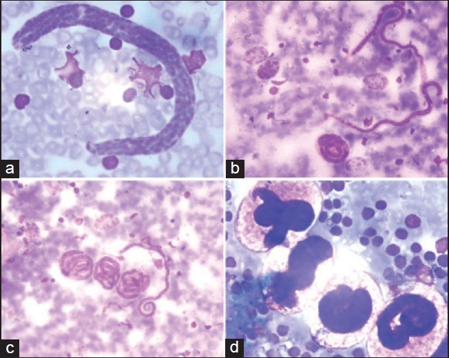
This was a case report of a 6-year-old male from eastern Uttar Pradesh presented with the complaints of axillary swelling, fever and loss of appetite. On examination, swelling was 3 cm × 2 cm in size, freely mobile, firm and non-tender. Overlying skin was normal and clinical diagnosis of tubercular etiology was made. Fine-needle aspiration (FNA) was done. Air-dried smears were stained with Giemsa stain. Smears showed many short and blunt larvae without any distinct sheath and nuclei. Numerous round to oval eggs with short coiled larvae inside them were also seen. No microfilariae with classical morphological features were seen [Figure 1]. A diagnosis of filarial lymphadenopathy was made.
 |
Figure 1: (a) Short and blunt larvae of filarial worm (Giemsa, ×1000) (b and c) Larvae and coiled filarial worm with early form of egg (Giemsa, ×400) (d) Embryonated eggs with coiled filarial worm (Giemsa, ×1000)
Click here to view |
| Discussion |
Lymphatic filariasis is transmitted by mosquitoes and is caused by closely related nematodes, Wuchereria bancrofti and Brugia species, which are responsible for 90% and 10% respectively. [6] The spectrum of diseases caused by filariasis include (1) asymptomatic microfilaremia, (2) recurrent lymphadenitis, (3) chronic lymphadenitis with swelling of the dependent limb or scrotum and (4) tropical pulmonary eosinophilia. [6] The major vectors are culex mosquitoes in urban areas and anopheles in rural areas. [2] The cytology of the filarial infestation can reveal microfilaria with or without adult worms and associated eosinophils, neutrophils and mononuclear cells. [4] Microfilaria may present in an unusual form even when filariasis is not suspected so careful screening of cytology smears help in making quick and efficient diagnosis of unsuspected cases of filariasis. [4] In present case, clinical diagnosis was of tubercular origin and FNAC plays a significant role in correct diagnosis of these lesions.
| Conclusion |
The case was considered worth documentation to highlight the finding of filarial eggs in FNA of lymph nodes, which can be missed or misdiagnosed by an inexperienced pathologist leading to delayed or wrong treatment of a curable disease and one should always consider the filariasis in the differential diagnosis of lymph node swelling, especially in endemic areas.
| References |
| 1. | Kapila K, Verma K. Gravid adult female worms of Wuchereria bancrofti in fine needle aspirates of soft tissue swellings. Report of three cases. Acta Cytol 1989;33:390-2. |
| 2. | Park JE, Park K. Textbook of Preventive and Social Medicine. 18 th ed. Jabalpur, India: M/S Banarasidas Bhanot Publishers; 2005. |
| 3. | Dreyer G, Addiss D, Roberts J, Norões J. Progression of lymphatic vessel dilatation in the presence of living adult Wuchereria bancrofti. Trans R Soc Trop Med Hyg 2002;96:157-61. |
| 4. | Hanmante RD, Mane UW, Vishwapriya, Suvernakar. An unusual presentation of filariasis as upper arm cystic swelling – Cytological diagnosis: A rare case report. Int J Recent Trends Sci Technol 2011;1:86-90. |
| 5. | Sivaselvam S. Role of fine needle aspiration cytology (FNAC) in the detection of microfilariae: A report of two cases and review of literature. Malays J Med Sci 2006;13:104. |
| 6. | McADam AJ, Sharpe AH. Infectious diseases. In: Kumar V, Abbas AK, Fausto N, Aster JC, editors. Robbin′s and Cotran Pathologic Basis of Disease. 8 th ed. Philadelphia: Saunders Elsevier; 2010. p. 395. |
Source of Support: None, Conflict of Interest: None
| Check |
DOI: 10.4103/1755-6783.133745
| Figures |
[Figure 1]


